We get the most knowledge of our surroundings through our eyes, which convey more information than any of our other senses. Every second the eyes absorb 10 million pieces of information and pass it on to the brain. Our vision adapts to different light conditions at cyber speed, refocusing within fractions of a second and distinguishing colors of several million shades.
Structure of the Eye
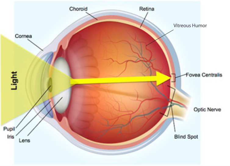
The eyes work much like a camera. Light passes through several parts of the eye—the cornea, anterior chamber, pupil, lens, and vitreous body before reaching the retina. The retina detects the light and converts it into signals. These signals travel through the optic nerve to the visual center at the back of the brain, where the brain processes them and creates the images we see.
Cornea
The cornea is the eye’s clear outer window. Light enters the eye through it before reaching the retina. It is made of tightly woven collagen fibers that form a transparent, dome-shaped surface. The cornea is just over half a millimeter thick and is covered by a thin layer of tear fluid. It also helps focus incoming light so that a clear image can form on the retina.
Pupil
The pupil is the opening in the center of the iris, the colored part of the eye. It adjusts its size to control how much light enters the eye. In bright light, it becomes smaller to protect the eye from excess light. In darkness, it widens to let in as much light as possible. The pupil can also change size in response to emotions such as fear, excitement, or joy.
Iris
When you look into someone’s eyes, you can see the iris as a colored ring surrounding the black pupil. The iris blocks light and contains two muscles that control the pupil’s size. These muscles make the pupil smaller or larger depending on the amount of light. The color of the iris is determined by pigments and inherited genes. Its tiny spots, lines, and patterns begin forming before birth. Every iris pattern is unique, even identical twins have different patterns.
Lens
The eye’s lens (lens crystallina) is a transparent, converging lens. It focuses the light entering through the pupil to form a sharp image on the retina. The lens is flexible and can change its shape to focus on objects at different distances. A small muscle, called the ciliary muscle, controls these changes. This adjustment is known as accommodation. As people age, the lens gradually becomes less flexible. This makes it harder to focus on nearby objects, a condition commonly known as presbyopia.
The Retina
The retina is the light-sensitive layer at the back of the eye. It contains about 127 million light receptors, which detect light after it passes through the cornea, lens, and vitreous body. These receptors convert light into nerve signals that can be sent to the brain. There are two main types of light receptors: cones, which allow us to see colors, and rods, which help us see differences between light and dark, especially in dim conditions.
The Optic Nerve
The optic nerve carries information from the retina to the brain. It is a thick bundle of nerve fibers, about half a centimeter wide. The optic nerve leaves the eye through a point on the retina called the optic disc. This area is also known as the blind spot because it contains no light receptors. As a result, a tiny part of each image is not detected. We usually do not notice this because the brain fills in the missing information.
Why is this relevant for stroke survivors with visual field deficits?
Approximately 30% of stroke survivors experience a visual field deficit and for 20% this deficit is permanent. Understanding the eye is important as damage to the visual pathway (often following a stroke or head trauma) and the consequent vision disorders and visual field deficits may result in a substantial difference to one’s daily life.
Visual field loss in the central visual field is of particular importance. Depending on the severity of the disorder, all aspects of everyday life, for example reading, eating, dressing or watching television may become difficult. It is imperative that anyone who experiences a neurologically induced visual field deficit reach out to seek dedicated specialist care to address these visual impairments as soon as practicable.
If you want to know more about vision problems after stroke or brain injury and available treatments, you can visit our informative website here.