


Blind areas in the eye can be the result of an eye disease, vision disorders or a visual field loss – the “blind spot” however is an absolutely normal phenomenon found in every human being.
The picture below shows the visual impression of a healthy left eye – why is there a black circle shown just to the left of the center? This is the part the healthy left eye does not see – the blind spot that everyone has in their left and right eyes. You might say: “No, not me, I have no blind area in the eyes, I can see everything.” We will show you in this blog that even with completely healthy vision, this is not true!
What is the blind spot?
The inside of every eye is lined with the retina, which maps everything that we see as an overall picture dot by dot, and converts it into electrical signals. These signals are relayed to the brain via nerve fibers. All the nerve fibers of the retina come together at a defined position in the eye and create a nerve bundle. This bundle progresses to the brain in form of the so-called optic nerve where the signals are further processed. Only then we “see”. At this defined position in the brain, at the point where the optic nerve connects the eye to the brain, there is no retina. It is at this position that part of the overall picture is not mapped and therefore is not seen; at this moment we are effectively blind, this is the “blind spot”.
This is best explained with pictures:
Image 1: Diagram of a human eye with retina, optic nerve and blind spot
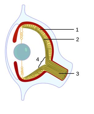
©Caerbannog via Wikimedia Commons (https://commons.wikimedia.org/wiki/File:Evolution_eye.svg)
1. Retina 2. Nerve fibers 3. Optic nerve 4. Blind spot
Image 2: Half model of the back of the eye, showing the retina, sustentative blood vessels and the entrance of the optic nerve (shown in white)

© “Wissen macht Ah!“
The retina covers the inner back half of the eye and consists of cones (responsible for color vision) and rods (light-dark-vision). During a baby’s development in the womb the retina is created as an immediate protuberance of the brain, therefore the light-sensing cells are directed to the inner side of the head. Therefore the nerve fibers that direct the information from the light-sensing cell to the brain are in the inner side of the eye. To be able to transport the information to the brain, the nerve fibers have to break through the retina. This is done by the optic nerve bundle at a certain point, the blind spot, about 15 degrees peripherally (in the left eye to the peripheral left, in the right eye to the peripheral right).
Because the position of the blind spot is different in each eye the overall picture is seen by the respective other eye to make the picture whole.
You will now say: “Still, I would have to have a blind area, e.g. when I cover the right eye and only see with my left eye. But there is none.” Well, you have not considered the trickiness of our brain. It extrapolates the most probable picture in this area from the visual information at the borders of the blind spot and simply fills it in. This part of the picture is self-created.
Since this is a very small area, and since most of the time we see the overall picture with both eyes, it is highly unlikely that we miss an important part of our surroundings through the blind spot. However, there are some nice little visual experiments that can demonstrate the existence of the blind spot quite strikingly.
Experiment 1
Please look at the detail of this picture. On the left side you see three blue birds, on the right side one red bird.

Now please cover your left eye.
With the right eye fixate on the left blue bird and move your head slowly towards the screen and then back again.
Did you notice? At a certain position the red bird suddenly disappears!
The distance to the screen is dependent on the size of your screen. The larger the screen, the further back you have to move your head. Just try it!
Only when you “outwit” eye and brain, as in this trial, you can make the blind spot “visible“.
Experiment 2
Look this charming gentleman straight in the eyes, cover your right eye and move your head towards the screen and back again; you will find that the coin suddenly disappears, like magic!
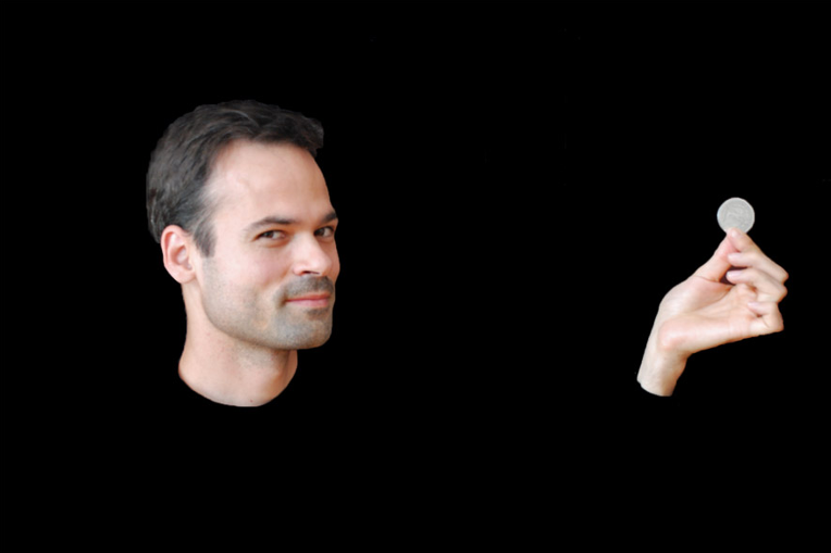
https://zaubereihoch3.files.wordpress.com/2011/07/blinder-fleck.jpg
Blind spots and neurological damage
Blind spots are a normal aspect of our vision and do not cause any visual problems. A stroke or other brain damage often causes damage to the visual pathway in the brain, and this can cause much larger blind areas in our vision – homonymous hemianopia, quandrantanopia or a scotoma. These can lead to severe impairment of vision and have an impact on everyday living. NovaVision’s NeuroEyeCoach and Vision Restoration Therapy are specifically designed to help people who have had a stroke or other brain damage and who, as a result, have a visual defect. Contact us for more information.
Written by: Jessica Einwiller
Resources: from 28.06.2016
Picture.: 1http://www.wdr.de/tv/wissenmachtah/bibliothek/blinderfleck.php5
https://de.wikipedia.org/wiki/Auge
