

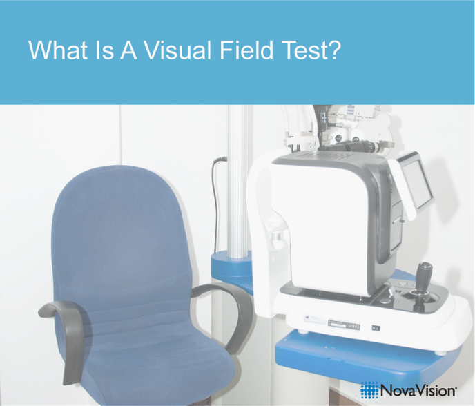
 When you have problems with your vision and go to an ophthalmology practice, often not only your eyes and their visual acuity are measured, but also your visual field is examined with a perimeter.
First of all: What is the visual field?
The visual field is the area of your vision that you can perceive when you look straight ahead without moving your eyes or head. Within the visual field our visual quality is different, depending on the area- in the center of our view, where we fixate objects, text, faces etc. with our eyes, we see particularly sharp. Further to the sides we see more blurry (peripheral vision). In the periphery it is most important to notice movements, so that eyes and head can be moved into that direction.
What happens during a perimetry (visual field test with perimeter)?
The classic perimeter measures the visual field using a hemisphere shaped bowl (hollow sphere) presenting light stimuli on its inner surface. The examined person sits in a darkened room and puts the head on a chinrest within the hemisphere. Every light stimulus seen is to be confirmed by pressing a response key. Mostly the examination is done monocularly, that means one eye is covered at a time to test the visual field of each eye separately.
Static and kinetic perimetry need to be differentiated. During kinetic perimetry, light stimuli are moved into the visual field. The examined person shall press a response key when a light stimulus is perceived. This method determined the outer limits of the visual field.
During static perimetry, stationary light stimuli are presented with varying brightness levels at different positions throughout the visual field. For every position the brightness level needed to see the light stimulus is determined. The overall result shows a visual performance profile for all areas of the tested visual field.
Depending on the test program selected (e.g. 90 degree visual field test, or 30 degree central field test only) the examination may take between 7 and 15 min.
During a visual field examination it is particularly important not to move the head or the eyes. The person examined is required to always look at a certain spot in the perimeter, in other words fixate a fixation point. This spot could as an example, be in the center of a circle of red light dots. During the examination, white light will flash intermittently at this center location which requires a response by pressing a button. The examiner observes the eye enlarged through a camera and is able to see eye movements instantly.
In addition to the fixation task, light dots are presented at other locations in the hollow sphere that also require a response by pressing a button. Because at each position light dots are displayed several times with different brightness levels, it is possible to determine a threshold, the minimal brightness needed to detect the light at this position.
The examination is designed in a way that even with perfect vision one will not be able to detect every light dot at the minimal brightness level. Vision decreased with higher age, so that brighter light is needed for detection. Eye or brain diseases can result in impairments or loss of vision in the whole visual field or in parts of the visual field. The pattern of visual field loss can provide important information about the underlying disease. Here you may learn more about visual field defects resulting from neurological damage such as stroke.
Example of a perimetry results report:
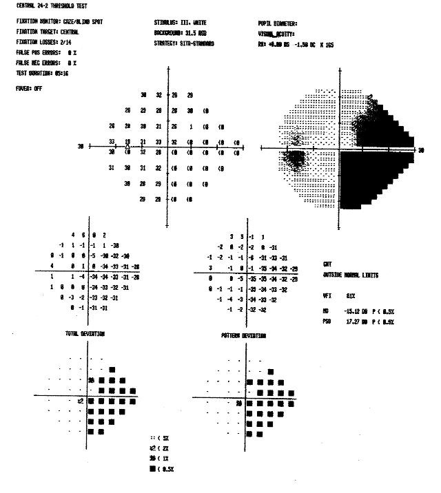 At the top of the report you find a description of the test parameters, e.g. which program (central 24-2 threshold test examining the central 24 degrees of the visual field), which stimulus size and color (size III, white) etc.
In this case the left eye was tested, the right eye was covered.
The values for “fixation losses“, for “false positive errors“ and “false negative errors” reflect the reliability of the examination:
- Fixation losses: how often was no response given when the white light flashed at the fixation point location? In this report 2/14, the light flashed 14 times, 2 times no response was given and fixation was lost.
- False positive errors: how often was the response button pressed, even though no light dot flashed? In this report 0 %, all responses were correctly given.
- False negative errors: how often was the response button NOT pressed when a bright light dot flashed, while there had already been a response to a less bright light dot at the same position? In this report 0 %, all responses were given as to be expected, and were correct.
In the below parameters you will find several graphical displays with different values:
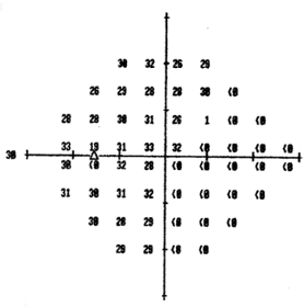 This display provides for every test position of the examination a number in “dB” scale, that is “Decibel”. This value is well-known with regard to sound volume, but is also a measuring unit for luminosity/brightness.The higher the value, the lower was the minimal brightness needed to detect the light dot at this position (and the better the visual performance level). In the central visual field the numbers are highest; this is the area of best visual acuity. Even with intact vision, the acuity decreases to the periphery. The term “<0”means that at this position even the brightest light dot did not elicit a response; no visual ability could be measured.
 In the second graph, the numerical values are translated into different patterns. The more light-colored the pattern, the better is the visual performance level.
On the right side of the graph is a large black area where there was no response to light dots. When the same black area is also found when testing the person’s right eye, this would be an indication of „incomplete homonymous hemianopia to the right“, a visual field loss resulting from neurological damage.
On the left side there is a small black circle. This does not reflect a vision impairment but marks the position of the blind spot in the left eye. You find it in every human being; here the optic nerve enters the eye, an area without retina which is indispensable for vision. In a perimetric examination the display of the blind spot is an indication of good fixation, and therefore of good result reliability.
The other four graphical displays of the overall report depict the visual field test outcome in relation to defined questions, for example deviation from healthy age matched controls.
Next week we describe the difference between a perimetry and our online visual field test.
You can take our online vision testvision test which is designed to help victims determine if they have experienced neurological vision loss after a stroke or brain injury and can be performed online with instant preliminary results. To properly determine the presence of a visual defect, the results of this free vision test should be combined with a complete vision evaluation with a medical professional.